Furosemide and Bone Health: Risks, Prevention, and What to Expect
Learn how furosemide affects calcium, PTH and bone density, who’s at risk, and practical steps to protect your skeleton while staying on the drug.
When talking about calcium loss, the gradual reduction of calcium stored in bones and teeth that weakens the skeletal framework, most people think of aging, but the story is far richer. Calcium loss is a type of mineral depletion that can start in your teens if diet or hormones are off‑balance. It doesn’t happen in isolation – it links directly to osteoporosis, a disease where bones become porous and fracture‑prone and to the body’s ability to maintain bone density, the measure of mineral content in each cubic centimeter of bone.
One of the biggest drivers behind calcium loss is vitamin D deficiency, a condition that hampers intestinal calcium absorption. Without enough vitamin D, even a diet rich in dairy can’t replenish skeletal stores, leaving the parathyroid glands to overproduce parathyroid hormone (PTH), a hormone that pulls calcium from bone to keep blood levels stable. That hormonal tug‑of‑war is a classic example of the semantic triple: "vitamin D deficiency influences calcium loss" and "parathyroid hormone drives bone demineralization".
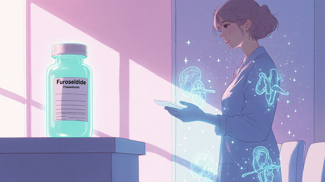
Beyond nutrition and hormones, lifestyle choices shape how quickly calcium drains from your framework. Long periods of inactivity, especially weight‑bearing exercise, mean less mechanical stress on bone, which reduces the stimulus for new bone formation. Smoking and excessive alcohol act like chemical thieves, accelerating osteoclast activity that chips away at calcium stores. Even certain medications—corticosteroids, loop diuretics, and some anticonvulsants—can tip the balance toward loss, a direct link showing that "medication use contributes to calcium loss".
Age‑related changes also matter. After 30, the body’s ability to synthesize vitamin D from sunlight declines, and the kidneys become less efficient at converting it to its active form. At the same time, intestinal calcium absorption drops by up to 1% per year. These physiological shifts create a cascade: reduced vitamin D, higher PTH, and faster bone turnover, which together form the semantic chain "aging reduces vitamin D, leading to increased parathyroid hormone, resulting in calcium loss".
When calcium loss slips into the clinical realm, doctors measure it with a DEXA scan that reports bone mineral density (BMD). A T‑score below –2.5 signals osteoporosis, the most severe stage of calcium depletion. However, even a T‑score between –1.0 and –2.5 indicates osteopenia, a warning zone where intervention can halt further loss. Knowing your BMD helps you see where you stand on the calcium loss spectrum and choose the right prevention plan.
Nutrition remains the first line of defense. Foods high in calcium—dairy, leafy greens, fortified plant milks—provide the raw material, while vitamin D‑rich sources like fatty fish, egg yolks, and sunlight exposure act as the catalyst. For many, a supplement of 800–1,200 mg calcium and 800–1,000 IU vitamin D daily fills gaps, especially in winter months. Pairing calcium intake with a small amount of vitamin K2 supports proper placement of calcium into bone rather than arteries, a subtle but important nuance.
Exercise, particularly resistance training and high‑impact activities like jumping or brisk walking, sends mechanical signals that tell bone‑building cells (osteoblasts) to lay down new calcium. A routine of three 30‑minute sessions per week can offset up to 10% of age‑related loss. Even simple habits like taking stairs, carrying groceries, or doing body‑weight squats keep the skeleton engaged.
Medical interventions become necessary when lifestyle tweaks aren’t enough. Bisphosphonates, denosumab, and selective estrogen receptor modulators (SERMs) are drugs that slow osteoclast activity, effectively reducing calcium loss. For severe cases, hormone replacement therapy or anabolic agents like teriparatide can actually stimulate new bone formation, a reversal of the usual calcium‑draining process.
Monitoring is key. Regular blood tests check calcium, vitamin D, and PTH levels, while repeat DEXA scans every 1–2 years track bone density trends. Early detection of rising PTH or falling vitamin D gives you a chance to adjust diet, supplements, or medication before noticeable fractures occur.
Putting it all together, calcium loss is a multifactorial issue that intertwines nutrition, hormones, lifestyle, and age. Recognizing the connections—vitamin D deficiency influences PTH, which in turn drives bone demineralization, leading to osteoporosis—helps you target the right levers. Whether you’re looking to prevent the first sign of bone weakening or manage an existing condition, the strategies outlined above give you a roadmap.
Below you’ll find a curated set of articles that dive deeper into each of these areas. From how specific drugs affect calcium balance to practical tips for boosting bone health on a busy schedule, the collection offers actionable insights you can start using right away.
Learn how furosemide affects calcium, PTH and bone density, who’s at risk, and practical steps to protect your skeleton while staying on the drug.